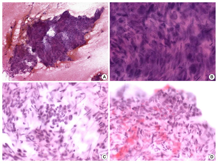
Fig. 2.
Cytologic features of metastatic leiomyosarcoma. (A) Hypercellular spindle cell clusters are arranged in a fascicular pattern. (B) The cells show indistinct cell borders, eosinophilic cytoplasm, and hyperchromatic, large, elongated nuclei with blunt ends. (C, D) Normal follicular cells are admixed with pleomorphic tumor cells (C) or discovered around tumor cell clusters (D).

