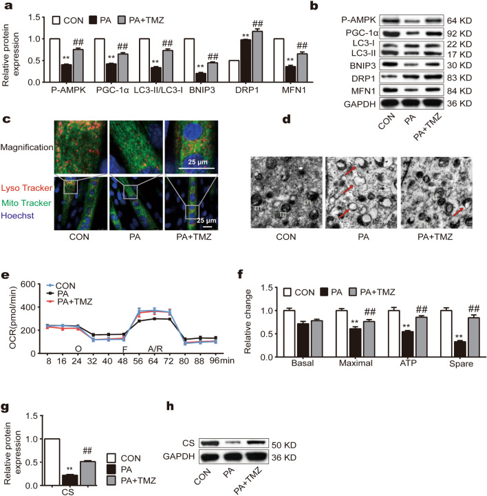
Figure 2.
TMZ promotes PA-attenuated mitochondrial quality control (MQC) and functions in muscle cells. (a, b) P-AMPK, PGC-1α, LC3II/LC3I, BNIP3, MFN1, and DRP1 protein expression were determined by Western blot. (c) Laser scanning confocal microscope by using LysoTracker and mitoTacker. The colocalizations of lysoTracker and mitoTracker represented mitolysosomes (scale bar = 25 μm). (d) Transmission electron microscope imaging. The damaged mitochondria were indicated in TEM images (red arrow). (e, f) OCR: cellular oxygen consumption. m: mitochondria. (g, h) CS protein expression was determined by Western blot. *, ** represented p < 0.05, p < 0.01 in comparison with the Con group, respectively; #, ## represented p < 0.05, p < 0.01 in comparison with the PA group, respectively.

