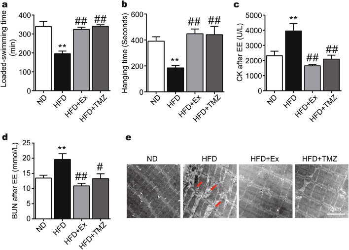
Figure 5.
TMZ regulates muscle function in the HFD mice comparably to exercise. (a) The forced weight-loaded swimming time was recorded as exercise capacity (EE exhaustive exercise). (b) The limb strength of the skeletal muscles was tested by the inverted screen test, meanwhile the fall-off time was recorded. (c, d) Plasma creatine kinase (CK) concentrations and plasma urea nitrogen (BUN) were observed after EE. (e) The skeletal structures of HFD mice were observed by transmission electron micrograph after EE with a scale of 2 μm. The red arrow represented the disordered and ruptured arrangement of the skeletal muscle fibers. *, ** represented p < 0.05, p < 0.01 in comparison with ND group, respectively; #, ## represented p < 0.05, p < 0.01 in comparison with HFD group, respectively.

