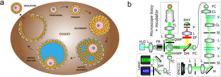
Fig. 1. Characterizing mechanical properties of ovaries ex vivo with Brillouin microscopy.
a Schematic of the developmental cycle of follicles during mouse folliculogenesis. A follicle consists of the oocyte (pink) with its nucleus (blue), surrounded by the somatic cells (orange) and theca cells (gray). The oocyte grows in size during transition to secondary follicle stage, followed by the emergence of fluid-filled lumen (blue) in the antral follicle stage. The oocyte is eventually released during ovulation, and upon fertilization undergoes embryo development. The interstitial tissues of the ovaries comprise of stromal cells, extracellular matrix, and vasculature. b Schematic of the confocal Brillouin imaging setup. L = lens, TL = tube lens, PMT = photomultiplier, P = pinhole, EF = emission filter, DM = dichroic mirror, FC = fiber coupler/collimator, QWP = quarter waveplate, HWP = half waveplate, PBS = polarizing beam splitter, S = shutter, FR = Faraday rotator, CL = cylindrical lens, V = VIPA, Sl = adjustable slit, V = VIPA, I = iris.

