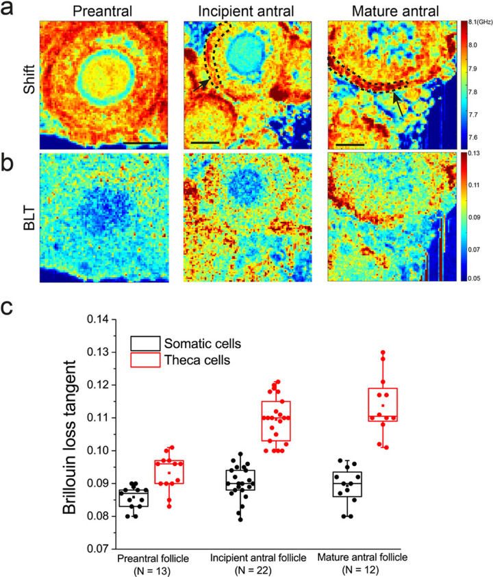
Fig. 3. Distinct mechanical compartments emerge during follicle maturation.
a Row shows representative maps of Brillouin shift for follicles at distinct stages of folliculogenesis. Scale bar = 40 μm. b Corresponding maps of BLT for a. c Boxplot of BLT for the outer theca cells versus the inner somatic cell layer of follicles at various stages of development. Each data point corresponds to the average signal of somatic or theca cells in one follicle. Black arrows indicate the follicles under consideration, black dashed lines indicate the region of interest for the theca cell layer.

