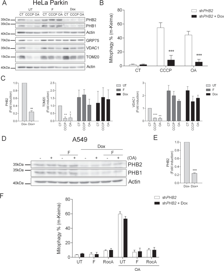
Fig. 5. Effect of PHB depletion in Parkin-dependent and -independent mitophagy.
HeLa Parkin shPHB2 cells (CT) were treated for 16 h with 10 μM CCCP or 1 μM oligomycin/1 μM antimycin A (OA) in the absence (UT) or presence of 10 μM fluorizoline (F), or after a 72 h 200 ng/mL doxycycline (Dox) treatment (A, C). HeLa Parkin shPHB2 cells (CT) were treated with 200 ng/mL doxycycline for 72 h and then treated with 10 μM CCCP or 1 μM oligomycin/1 μM antimycin A (OA) for 16 h (B). A549 shPHB2 cells (CT), previously treated or not with 200 ng/mL doxycycline (Dox) for 72 h, were treated with 1 μM oligomycin/1 μM antimycin A (OA) for 24 h in the absence (UT) or presence of 10 μM fluorizoline (F) or 500 nM rocaglamide A (RocA) (D, F). Protein levels from whole-cell lysates were analyzed by western blot and actin was used as a loading control. These are representative images of three independent experiments (A, D). m-Keima was measured by flow cytometry and referenced to their corresponding untreated controls and it is expressed as the mean ± SEM (n = 3 independent experiments) of the percentage of mitophagy positive cells (B, F). PHB2, TOM20, and VDAC1 protein expression levels were quantified. Mean ± SEM (n = 3 independent experiments) (C, E). ***p < 0.001 Dox-treated versus Dox-non treated cells (B); *p < 0.05, **p < 0.01, ***p < 0.001 CT versus CCCP and OA-treated cells (C, E).

