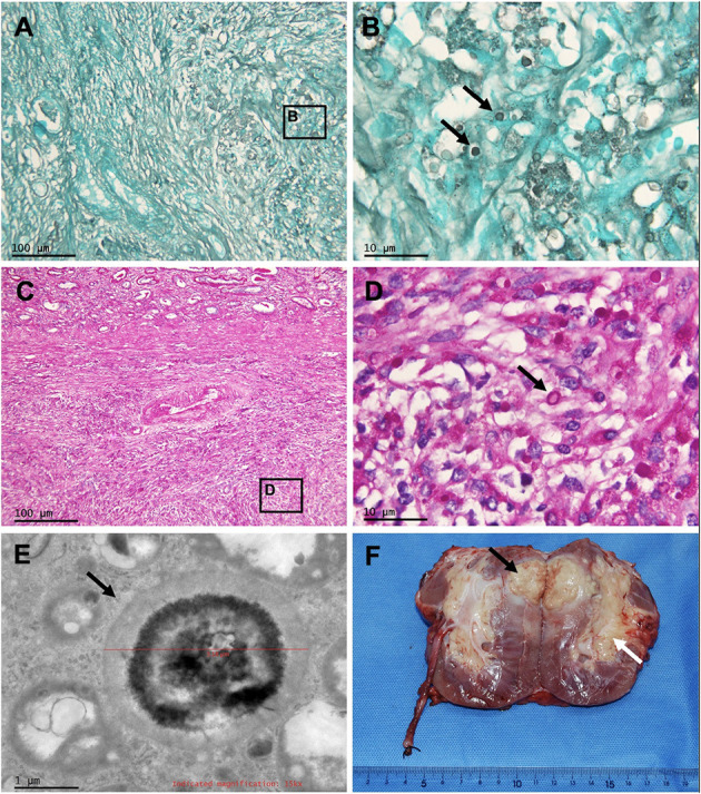
Figure 3.

Pathology of the masses of the transplanted kidney, and gross photograph of the allograft nephrectomy specimen. Periodic acid-Schiff staining and Grocott methenamine silver staining of renal mass at 100 × (A,C) and at 1,000 × (B,D) show granuloma caused by Cryptococcus (arrows). Electron microscopy of renal mass (E) shows the Cryptococcus (arrow). (F) Renal cryptococcoma (black arrow) and enlarged renal crptococcoma (white arrow).
