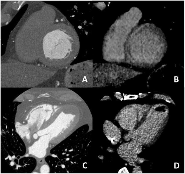
Figure 6.

In (A,B) an example of non-ischemic fibrosis on the interventricular septum is shown; more specifically, in (A) angiographic phase CT is presented with corresponding late CT acquisition well demonstrating non-ischemic mid-wall myocardial fibrosis in (B). In (C,D) a case of ischemic cardiomyopathy is presented; an apical left ventricular thrombus is evident both at first pass CT (C) and at late phase CT (D) from which the ischemic subendocardial fibrosis could be evidenced.
