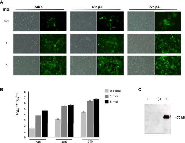
Figure 1.
BoHV-4-A-PPRV-H-ΔTK adaptation and characterization in ovine fetal lungs (OFLs). (A) Representative images of BoHV-4-EFGPΔTK-infected OFLs with different moi at 24, 48, and 72 h post-infection. Magnification, ×10 (all panels). (B) Viral titer was measured and expressed as Log10 TCID50 per ml of viral particles released at 24, 48, and 72 h post-infection when infected with 0.1, 1, or 5 moi, respectively. Values shown are the means ± standard errors of three independent experiments. (C) Western immunoblotting of OFL cells, infected with BoHV-4-A-PPRV-H-ΔTK (lane 2) or the parental BoHV-4-AΔTK used as a negative control (lane 1). (L) Mass ladder. The lanes were loaded with the same amount of total protein.

