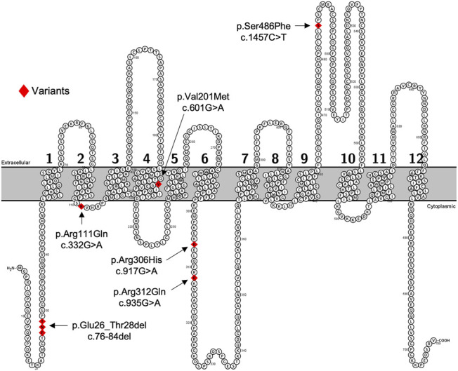
FIGURE 1.

Predicated 2-D structure of OATP2B1 full length transcriptional variant. Genetic variants of interest are highlighted in red and indicated by arrows with residue number and amino acid change. The predicted 2-dimensional membrane topology model of OATP2B1 was generated using Protter interactive protein visualization software (https://wlab.ethz.ch/protter/start/).
