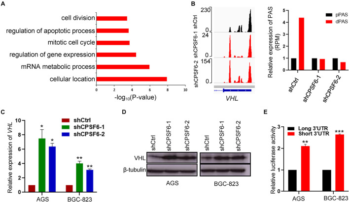
FIGURE 5.
CPSF6 downregulates the expression of VHL. (A) GO analysis of genes with shortened 3′UTR after knockdown of CPSF6 in AGS cells. (B) CPSF6 induced APA shift of VHL. Left, Integrative Genomics Viewer (IGV) genome browser exhibited the poly(A) site usage of VHL 3′UTR. Right, histogram showed the relative expression of the isoform with distal polyadenylation site (dPAS) relative to the one with proximal PAS (pPAS). (C) The mRNA levels of VHL in CPSF6 knockdown AGS and BGC-823 cells. (D) The protein levels of VHL in CPSF6 knockdown AGS and BGC-823 cells. (E) The luciferase reporter assay for the expression activity of the isoform of VHL 3′UTR in AGS and BGC-823 cells. *0.01 < p < 0.05; **0.001 < p < 0.01; ***p < 0.001).

