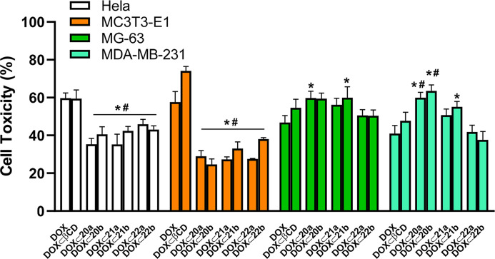
Figure 6.
Cytotoxicity of DOX and DOX inclusion complexes in different cell lines. HeLa, MC373-E1, MG-63, and MDA-MB-231 cells were incubated for 48 h in the presence of 1 μM DOX or an equivalent concentration of DOX occluded onto βCD, PEI-BP-CD, and PEI-MP-CD conjugates. The cell cytotoxicity (expressed as the percentage of the cell viability of the untreated cells minus the cell viability of treated cells) was determined by an MTT assay. The data are shown as mean ± SEM (n = 10). *p < 0.05 vs DOX-treated cells; #p < 0.05 vs DOX ⊂ βCD-treated cells.

