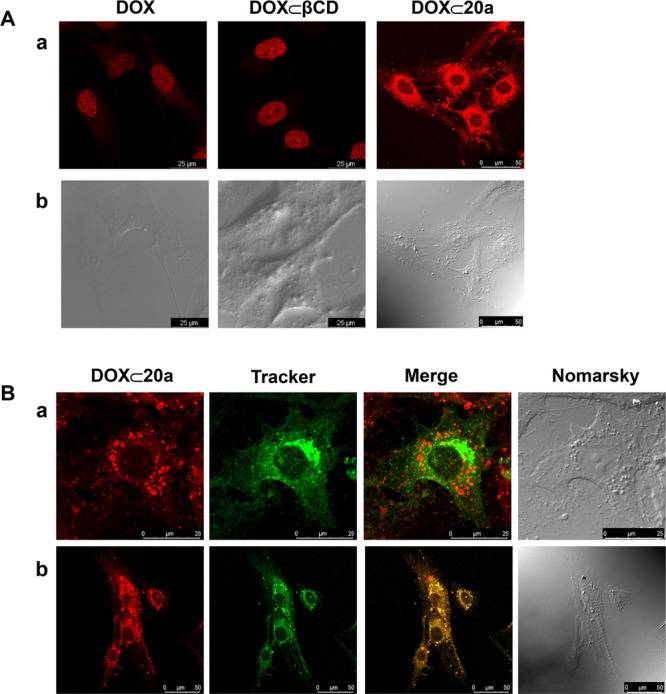
Figure 7.

Subcellular distribution of DOX ⊂ 20a. (A) MG-63 sarcoma cells were incubated for 2 h with DOX, DOX ⊂ βCD, or DOX ⊂ 20a, and then, confocal images were obtained (fluorescent (a) and Nomarski (b) images are shown). (B) MG-63 cells were preincubated for 30 min with either Alexa 488-labeled cholera toxin as a marker of late endosomes (a) or green mitotracker as a mitochondrial marker (b), and then, cells were incubated for 1 h with DOX ⊂ 23a, and sequential confocal fluorescence images were obtained.
