Figure 8.
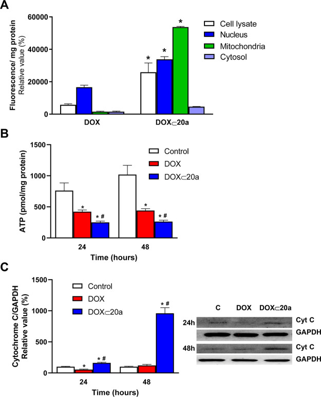
Mitochondrial targeting of the DOX ⊂ 20a inclusion complex. (A) MG-63 cells were incubated in the absence or presence of 1 μM DOX or equivalent concentration occluded in the DOX ⊂ 20a complex for 24 h. A subcellular fractionation of the MG-63 cells was carried out and fluorescence was measured in each fraction. Results are mean ± S.E.M. (n = 4). *p < 0.05 compared to DOX-treated cells. (B) MG-63 cells were treated under the same conditions as in A for 24 or 48 h, and the ATP concentration was determined in the mitochondrial fraction. (C) MG-63 cells were treated under the same conditions as in B, and the release of cytochrome c to the cytosol was measured by Western blot in the cytosolic fraction. Results are mean ± S.E.M. (n = 4). *p < 0.05 compared to untreated cells. #p < 0.05 compared to DOX-treated cells.

