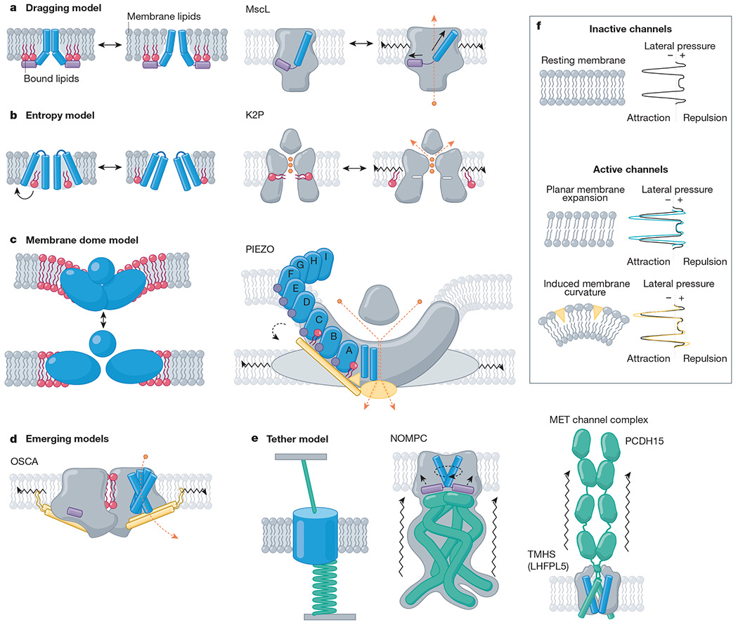
Fig. 2 |. Mechanistic models of mechanically activated ion channel gating.

Proposed mechanistic models with channel family examples. Amphipathic helices (violet), TM helices (blue), bound lipids (red), beam-like features (gold), tethers (emerald), ions (orange) and membrane lipids (grey) are indicated. a, Left, the dragging model. Lipids interact with an amphipathic helix and drag it outwards upon membrane expansion4. Right, MscL, for example, has an amphipathic helix on the internal leaflet (helix S1, violet) that drives a tilt to the pore-lining helix (TM1, blue) as it is ‘dragged’ outward under tension38. b, Left, entropy model. Lipids reside in hydrophobic pockets in the closed state and exit these pockets under membrane tension, inducing a conformational change132. Right, K2P, for example, has a fenestration occupied by lipid acyl tails (red) when inactive, whereas in active channels, this fenestration is closed and lipids are absent. c, Left, membrane dome model. Channel curvature within the membrane stores energy18. Right, PIEZOs, for example, expand and flatten134, gating the pore via interactions between the beam domain, the anchor domain and the CTD (gold)90. d, Emerging models. OSCA channels have lipid-occupied pores and an intersubunit cleft (red), an amphipathic helix (violet) on the inner leaflet, and a beam-like domain (gold) connected to pore-lining helices, which terminates a membrane-entrant hook domain (gold); all of these domains could have a role in gating23. e, Left, the tether model. Force is transmitted to the channel via a tether to the extracellular matrix, the cytoskeleton or both. For example, NOMPC (middle) is tethered to microtubules via its ankyrin repeat domain (emerald)112. Right, the MET channel complex is tethered to the neighbouring stereocilium via the tip link (PCDH15; emerald)16. f, Top, resting membrane tension. The transbilayer pressure profile reflects the lateral pressure experienced through the bilayer as a consequence of repulsion (positive pressure) of the lipid head groups, attraction (negative pressure) due to surface tension at the glycerol backbone, and steric hindrance (positive pressure) between the lipid tails32. Bottom, model membranes under tension. Planar membrane expansion thins the bilayer and increases the area occupied by each lipid. Membrane curvature is induced when suction is applied to the membrane or conical-shaped amphipathic compounds insert into the bilayer130,148.
