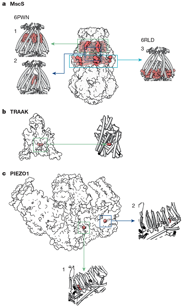
Fig. 3 |. Lipids observed in structures of mechanosensitive ion channels.

a, Lipids are observed in three locations in MscS structures. (1) one lipid per subunit is ‘hooked’ at the periplasmic leaflet12,13; (2) densities ascribed to lipid acyl chains reside inside the pore12,13 (PDB: 6PWN). (3) Two additional lipids per protomer are observed parallel to TM3b, below the membrane leaflet12 (PDB: 6RLD). b, In inactive structures of TRAAK (PDB: 4WWF), an acyl tail (green) occupies a fenestration below the selectivity filter62. c, Two lipid-like densities are observed in the PIEZO1 structure (PDB: 6BPZ): (1) in the region between the anchor domain and piezo repeat A, and (2) between piezo repeats B and C (second and third from the pore, respectively)19.
