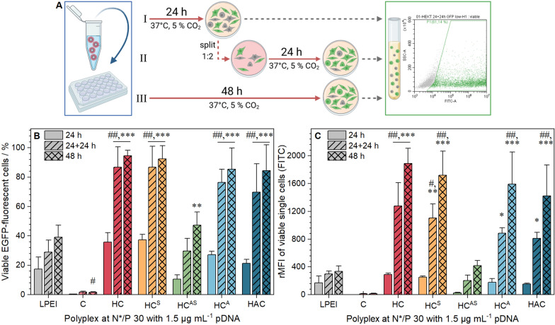
Fig. 6.
Transfection efficiency of polymers in HEK293T cells. EGFP expression of viable cells was analyzed via flow cytometry. A Schematic representation of the incubation method (created with BioRender.com). Cells were incubated with (layered) polyplexes of mEGFP-N1 pDNA and polymers at N*/P 30 in growth medium for 24 h (I), for 24 h followed by splitting of cells and medium and further incubation for 24 h (II), or for 48 h (III). Values represent mean ± SD (n = 3) of (B) viable, single EGFP positive cells (C) rMFI of all viable single cells relative to cells treated with polyplexes of pKMyc pDNA and polymers. ##Significant difference to HC-mic at respective time points (p < 0.001), */**/***significant difference to same polymer after 24 h (p < 0.05/0.01/0.001)

