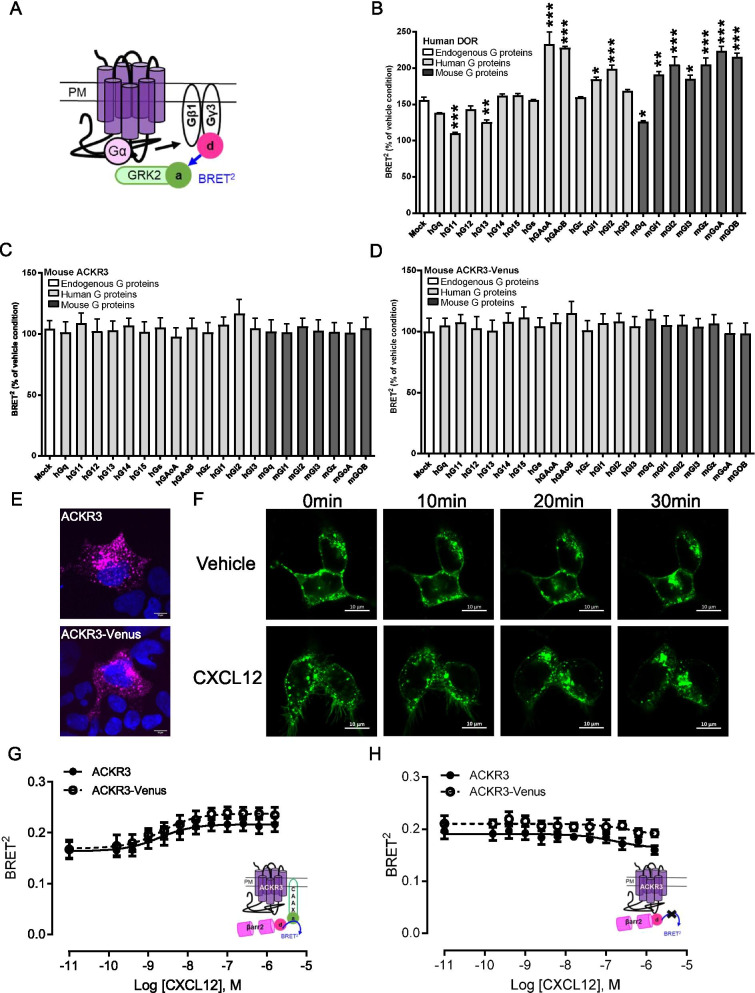
Fig. 1.
Functional characterization of ACKR3-Venus in HEK-293 cells. A Schematic representation of the BRET2 biosensor used to assess receptor induced G protein activation by monitoring the interaction between the dissociated hGγ3-RlucII (BRET2 sensor donor) upon activation and hGRK2-GFP10 (BRET2 sensor acceptor) [33, 78]. B–D HEK-293 cells were co-transfected with the indicated receptor (DOR, ACKR3 or ACKR3-venus), Gβ1, hGγ3-RlucII, hGRK2-GFP10 and the indicated human (h) or mouse (m) Gα subunits and then stimulated by agonist (see B and C, D below). Mock is a condition with cells not co-transfected with a Gα subunit encoding cDNA, thus detecting receptor-mediated activation of endogenous G proteins. Data are expressed as % of vehicle condition (Mean ± SEM, see Additional file 1: Figure S1A–C for vehicle conditions at each G protein), *p < 0.05, **p < 0.01, ***p < 0.0001 vs Mock condition, in 3 independent experiments. B G protein activation profile of the human Delta opioid receptor (DOR) following a 10 min stimulation with 30 µM of Met-Enkephalin. C, D G protein activation profile of the untagged (ACKR3) or Venus-tagged (ACKR3-Venus) mouse ACKR3 following a 10 min stimulation with 1 µM CXCL12. E Surface Flag ACKR3 (top) or Flag-ACKR3-Venus (bottom) receptors were labelled before fixation and permeabilization in HEK-293 cells to detect the surface pool of receptors only, using confocal microscopy. F HEK-293 cells were transfected with ACKR3-Venus and the time-dependent changes in the subcellular distribution of ACKR3-Venus was monitored using confocal microscopy in cells stimulated with 300 nM CXCL12 (bottom) and vehicle-treated control (top). G, H βarrestin2 recruitment assessed using the βarrestin2-RlucII/rGFP-CAAX ebBRET biosensor [34]. HEK-293 cells were transfected with either untagged or Venus-tagged mouse ACKR3 receptors, βarrestin2-RlucII, with (G) or without (H) the rGFP-CAAX BRET2 biosensor plasmid. Cells were stimulated for 10 min with increasing concentrations of CXCL12. Upon receptor activation, βarrestin2-RlucII (donor) is recruited to the plasma membrane (rGFP-CAAX, acceptor), resulting in an increase BRET2 signal. The LogEC50 values for the CXCL12-promoted βarrestin2 are 8.7 (CI 8.9–8.5) and 8.6 (CI 8.8–8.4); P = 0.96. The control experiment (H), with no rGFP-CAAX expression, is to rule out a potential contribution of the Venus tagged to ACKR3 to the ebBRET signal measured between βarrestin2-RlucII and rGFP-CAAX. Data are expressed as raw BRET signal (Mean ± SEM) from 3 independent experiments

