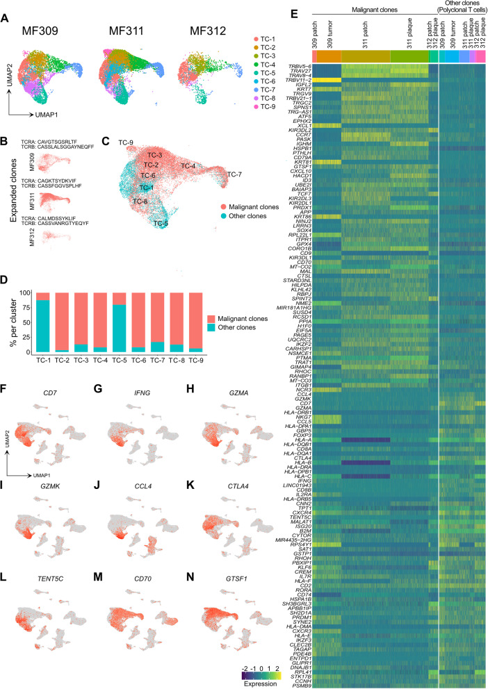
Fig. 2.
Characteristics of T cell clusters. A Separate UMAP plots of T cell clusters from skin samples of patients MF309, MF311 and MF312. B UMAP plots of the single top expanded clone (according to the most common α and/or β chain CDR3s aminoacid sequence) per patient. C Combined UMAP of T cell clusters colored according to clonality of TCR. Red: Cells with top expanded monoclonal TCR(s) from each sample; turquoise: cells with polyclonal TCR. D Percentage of monoclonal and polyclonal populations per T cell cluster. E Heat map displaying top differentially expressed genes between expanded clones and polyclonal TCR+ cells, according to the smallest adjusted p-value and average log fold change as calculated by logistic regression with Bonferroni correction; upregulation is indicated in yellow, and downregulation in blue/green; gene names are shown on the left. F-N Combined feature plots showing expression of selected genes differentially expressed between top expanded clones and the polyclonal infiltrate. Normalized expression level for each cell is color-coded (red) and overlaid onto UMAP plots. TC: T cells; UMAP: Uniform Manifold Approximation and Projection. TCR: T-cell receptor

