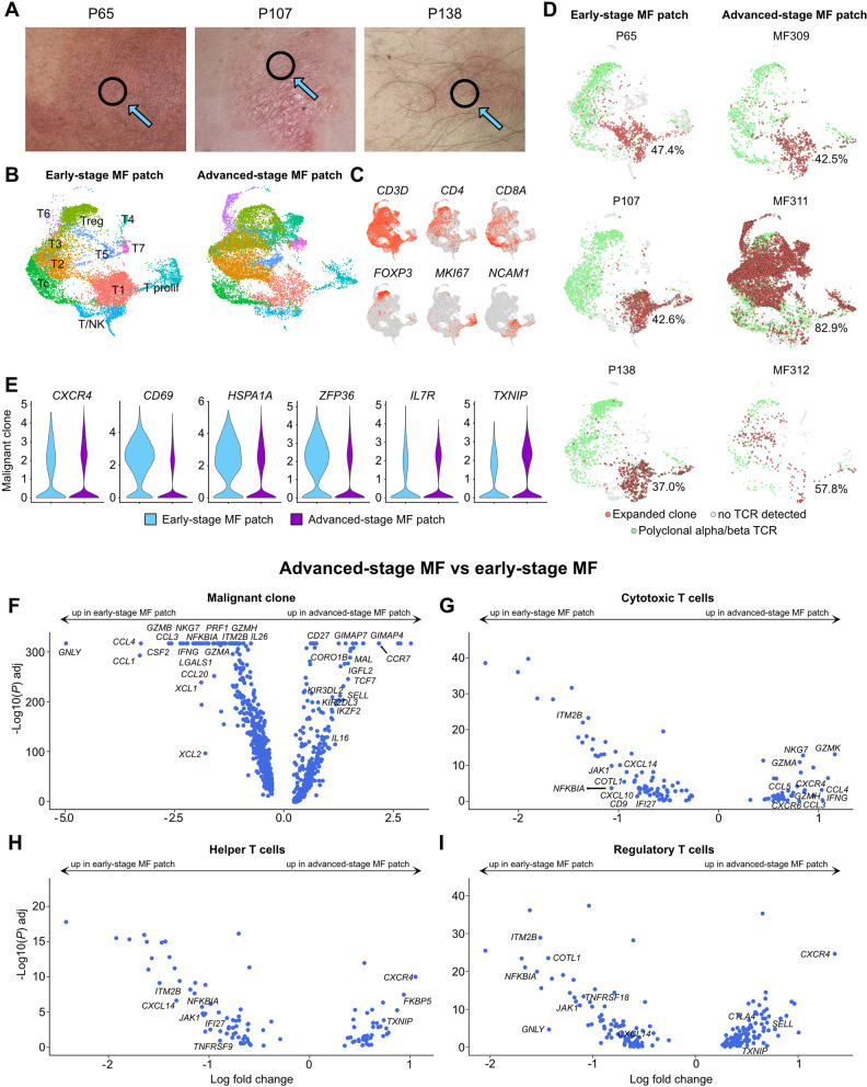
Fig. 6.
Comparison of patch lesions from advanced-stage vs. early-stage disease. A Pictures of patch lesions from 3 patients with longstanding, early-stage MF; black circles indicate biopsy location. B UMAP of 20,975 cells from three early-stage (P65, P107, P138) and three advanced-stage MF patches (MF309, MF311, MF312) according to similarity of their transcriptome. T-cell harboring clusters from Fig. S4 (clusters TC-1 to TC-8, Prolif and Treg, as depicted in Fig. S4A) were used for reclustering, resulting in 11 different color-coded clusters, split according to tissue of origin. C Feature plots showing expression of selected T and NK cell marker genes. Normalized expression level for each cell is color-coded (red) and overlaid onto UMAP plots. D UMAP plots of individual patients colored according to most common monoclonal (red) and polyclonal (green) αβ TCR; cells without TCR are displayed in grey. Percentages denote frequencies of malignant cells among all TCR+ cells for each plot. E Violin plots showing distribution of normalized gene expression levels in the top expanded αβ TCR clone in early (light blue) vs. advanced-stage MF (purple). F-I Volcano plot showing differentially expressed genes (DEGs) of the malignant clone, as well as polyclonal helper, cytotoxic and regulatory T cells between advanced and early MF patch lesions. Differential gene expression was defined as log fold change >∣0.25∣ calculated by logistic regression and Bonferroni correction. Regulatory T cells: FOXP3+ cells with polyclonal TCRs. Cytotoxic T cells: CD8A+ FOXP3- cells with polyclonal TCRs. Helper T cells: CD4+ FOXP3- cells with polyclonal TCRs. UMAP: Uniform Manifold Approximation and Projection

