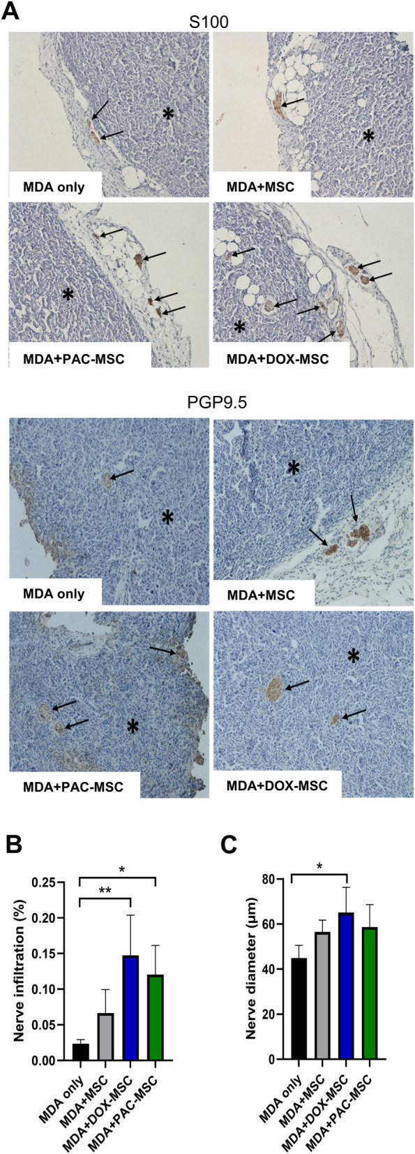
Fig. 4.

Chemotherapy-exposed MSC and nerve infiltration in tumor xenografts. (A) Two different polyclonal antibodies identifying nerve tissue (against S100 protein and PGP9.5 protein) stained nerve structures cross-section area in the tumor (indicated by arrows) and in the adjacent adipose tissue up to 2 mm from the tumor surface. (B) Nerve infiltration was expressed as percentage of the tumor section area in each experimental group. Increased nerve infiltration of tumors was observed in co-cultures with DOX-/PAC-MSC. (C) DOX-MSC co-injection also increased nerve diameter of detected tumors. For statistical analysis, one-way ANOVA with Tukey’s multiple comparisons test was used. Statistically significant results are highlighted with asterisks at p < 0.05 (*), p < 0.01 (**)
