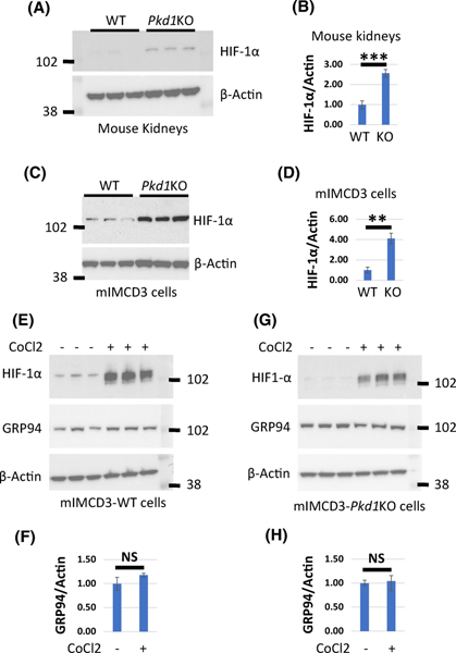
Figure 6. HIF-1α does not induce GRP94 expression in Pkd1KO and WT cells.
(A) Western blot expression of HIF-1α in three WT and three Pkd1-CKO-P20 kidneys. (B) Quantification of HIF-1α expression in (A). Protein levels in WT cells was set to 1. (C) Western blots showing HIF-1α levels in mIMCD3-WT and mIMCD3-Pkd1KO cells. (D) Quantification of HIF-1α expression in (C). HIF-1α level in WT cells was set to 1. (E and G) Western blots showing HIF-1α and GRP94 levels in mIMCD3-WT and mIMCD3-Pkd1KO cells treated with cobalt(II) chloride (CoCl2) as indicated. (F and H) Quantification of GRP94 in E and G, respectively. GRP94 level in untreated cells was set to 1. The bars represent mean ± SEM. **p < 0.01, ***p < 0.001, NS - not significant.

