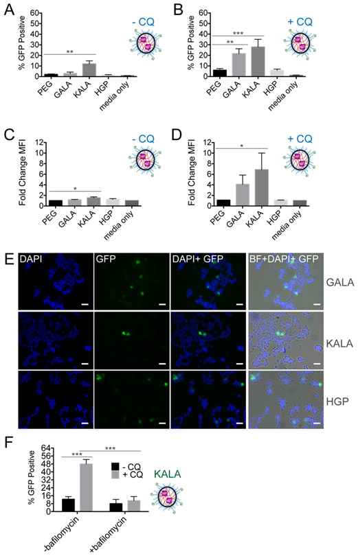
Figure 5. NG functionalization with pH-sensitive peptides influences endosomal escape.
(A) SEE after 24-hour incubation with surface modified PEG-PDS NG complexes at 0.75 mg/mL polymer concentration in HEKs1-10 cells and (B) In the presence of 80 μM CQ. SEM error bars pertain to individual biological replicates from independent NG batches, analyzed on different days. (C) SEE represented as change in GFP mean fluorescent intensity (MFI) of cells in CQ-free media. The change in MFI is generated after normalizing GFP MFI to the untreated, viable single-cell HEKs1-10 population. (D) In the presence of 80 μM CQ. (E) Visualized CQ-liberated endosomes after 24-hour incubation with the different pH-responsive-NG (20X fluorescent microscope objective: BF (bright-field), DAPI (nucleus stain, 405 nm), GFP (reassembly of C3KO-11 and GFP1-10, 488 nm)). SEM error bars pertain to four individual biological replicates from independent NG batches analyzed on different days. (F) SEE of KALA-PEG-PDS NG, with and without CQ and bafilomycin, an endosomal acidification inhibitor. For (A-D), one-way ANOVA performed against PEG-PDS where *p < 0.05; **p < 0.01; ***p <0.001; ****p < 0.0001. If no label present, no significant (n.s.) differences were found. For (F), individual unpaired t-tests were performed.

