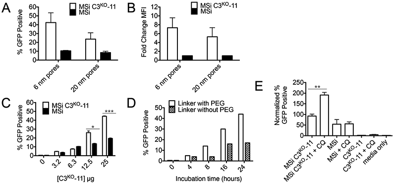
Figure 6. Detecting cytosolic delivery of C3KO-11 via mesoporous silica (MSi) nanoparticles by SEE.

(A) Influence of MSi pore size on SEE after 24-hour incubation with MSi complexes in HEK293T cells transfected with GFP1-10 and (B) Change in MFI. (C) Dose dependence of SEE after 24-hour incubation with MSi complexes in HEKs1-10 cells. (D) Effect of incubation time on SEE. (E) SEE improvement of various MSi complexes in the presence of 100 μM CQ. For (C) two-way ANOVA performed and for (E) unpaired t-test (two-tailed) executed where *p < 0.05; **p < 0.01; ***p <0.001; ****p < 0.0001. If no label present, no significant (n.s.) differences were found. For (F), individual unpaired t-tests were performed.
