
Figure 16.
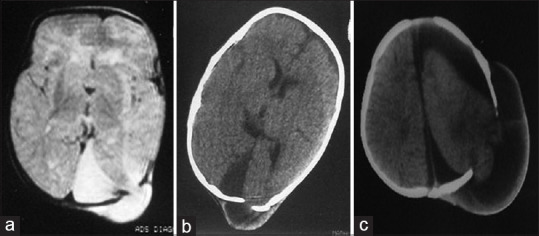
T2 axial magnetic resonance imaging (a), axial computed tomography (b and c) showing progressive cerebral migration and displacement
Official websites use .gov
A
.gov website belongs to an official
government organization in the United States.
Secure .gov websites use HTTPS
A lock (
) or https:// means you've safely
connected to the .gov website. Share sensitive
information only on official, secure websites.
T2 axial magnetic resonance imaging (a), axial computed tomography (b and c) showing progressive cerebral migration and displacement