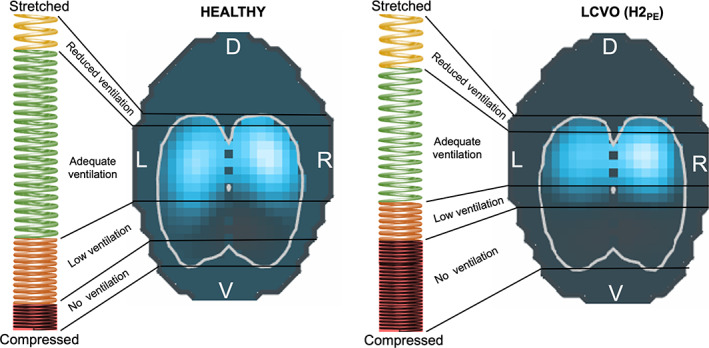
FIGURE 5.

The “spring model” illustrating the slinky effect of gravity on the distribution of ventilation. The differences in the coil stretch of the spring and the EIT image between a healthy horse (left) and a horse with left‐sided cardiac volume overload (LCVO; right) are displayed. The vertically oriented spring is stretched dorsally and compressed at the bottom. The corresponding EIT image demonstrates the areas of detected impedance change in light blue. An increase in extravascular lung water (right) leads to a greater number of coils in the dependent portion of the spring, which is analogous to a greater density of the lung tissue and therefore less ventilation detected in the EIT image
