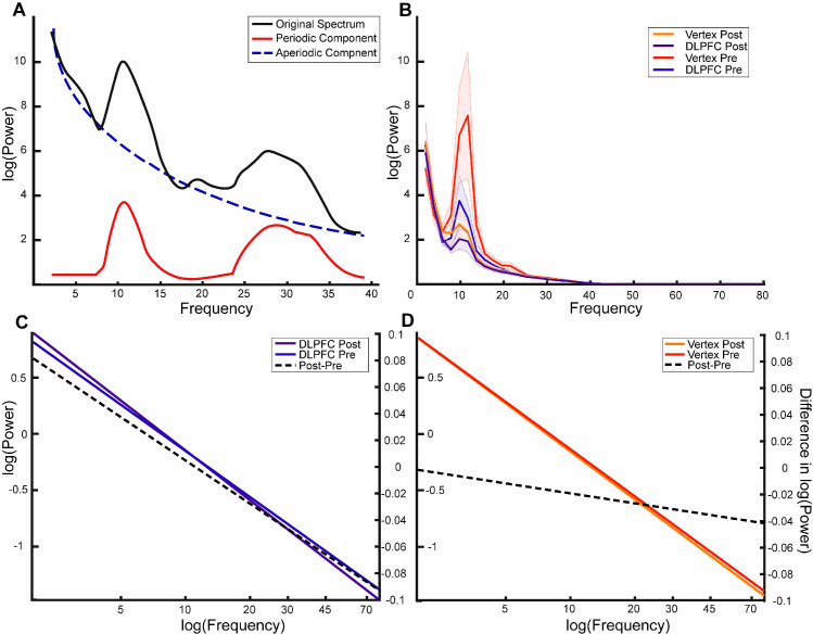
Fig 4. FOOOF analysis shows that beta effects are nonoscillatory in nature.
(A) Schematic representation of the different components in a given power spectrum. The black line represents a typical power spectrum that is to be separated. The blue line is the corresponding log function following removal of the periodic peaks, thereby representing aperiodic properties of the signal. (B) Power spectra separated by each condition. Shaded area indicates standard error. (C, D) Line plots of the mean aperiodic component before and after item presentation for the DLPFC and vertex condition, respectively. The right axis relates to the plotted post–pre difference (dotted line). The x-axis has been extended for illustrative purposes to highlight the differences in slopes between the difference conditions. The actual fit was performed on data in the 1–40 Hz range. The data and scripts used to generate this figure can be found at https://osf.io/dyxjv/. DLPFC, dorsolateral prefrontal cortex.

