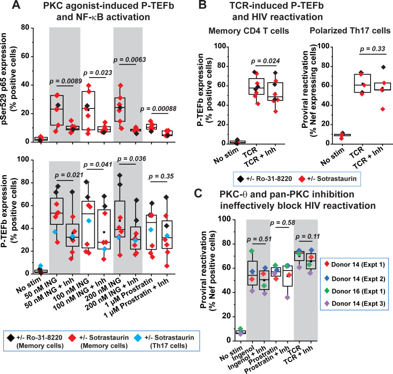
Fig 5. Coordinate reactivation of P-TEFb and latent HIV in primary T cells is largely independent of PKC activity.
(A) PKC-θ and pan-PKC inhibition are inefficient at repressing ingenol- or prostratin-induced expression of P-TEFb, but effective at blocking the activating phosphorylation of the p65 subunit of NF-κB at Ser529 in response to these phorbol esters. CD4+ memory T cells from four different donors and in vitro polarized primary Th17 cells were treated for 24 h with the indicated concentrations of either ingenol or prostratin in the presence or absence of either 100 nM sotrastaurin or 100 nM Ro-31-8220 prior to subjecting cells to dual immunofluorescence staining for pSer529 p65 (top graph) or for CycT1 and pSer175 CDK9 (bottom graph). (B) PKC inhibitors modestly repress TCR-mediated expression of P-TEFb in CD4+ memory T cells but are unable to block reactivation of proviral HIV in primary Th17 cells. Left graph; Measurement of P-TEFb expression by dual CycT1 and pSer175 CDK9 immunofluorescence staining of memory T cells that were TCR-activated with anti-CD3/anti-CD28 Dynabeads for 24 h in the presence or absence of treatment with either 100 nM sotrastaurin or 100 nM Ro-31-8220. Right graph; Assessment of the effect of PKC inhibition on proviral reactivation in ex vivo HIV-infected primary Th17 cells prepared from naïve CD4+ T cells belonging to three healthy donors. Latently infected cells were treated or not for 30 min with 100 nM Ro-31-8220 in two experiments or with 100 nM Sotrastaurin in three experiments prior to TCR co-stimulation with anti-CD3/anti-CD28 Dynabeads for 24 h. Thereafter, cells were analyzed by flow cytometry following immunostaining using a fluorophore-conjugated antibody towards HIV Nef. (C) PKC inhibitors are unable to repress the reactivation of latent HIV in primary Th17 cells that is in response to PKC agonists. Latently infected Th17 cells were treated or not for 30 min with either a combination of Ro-31-8220 and Gö 6983 (Donor 14 (Expt 1) and Donor 14 (Expt 2)) at 100 nM each or 100 nM Sotrastaurin (Donor 16 (Expt 1) and Donor 14 (Expt 3)) prior to TCR co-stimulation or challenge with 50 nM ingenol or 1 μM prostratin. Thereafter, cells were immunostained using a fluorophore-conjugated antibody towards HIV Nef and analyzed by flow cytometry. Statistical significance (p values) in A, B and C was calculated using a two-tailed Student’s t test.

