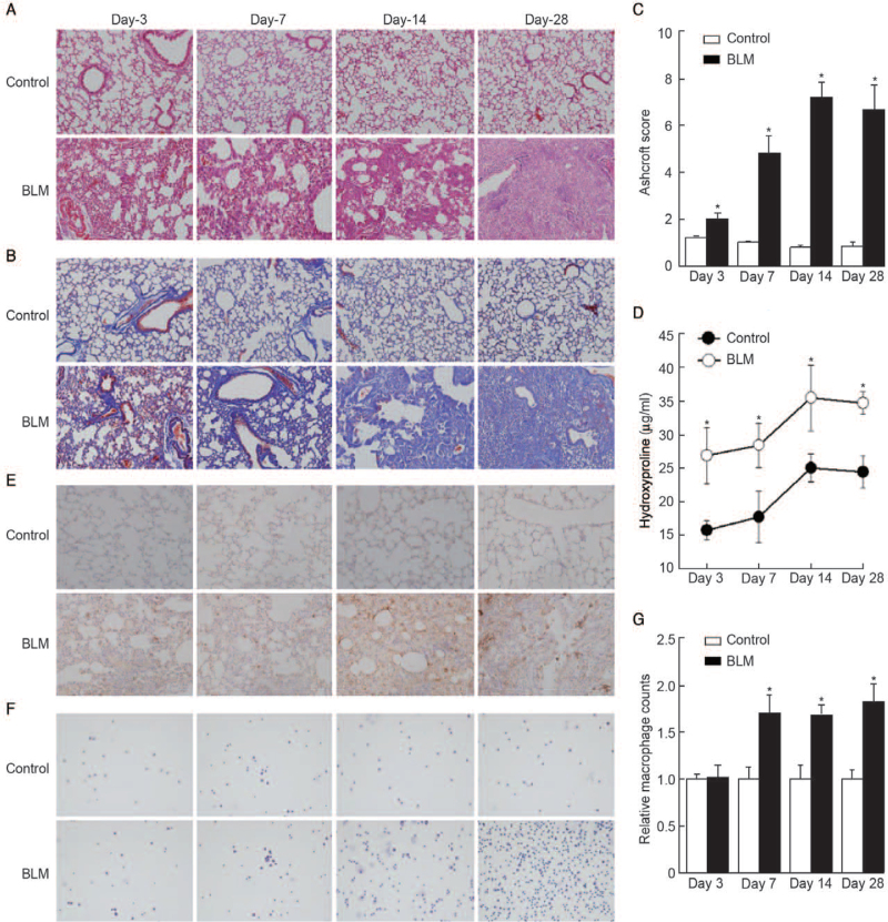
Figure 2.
Macrophage infiltration correlates with lung fibrosis. Representative microphotographs of lung sections from control and BLM (sacrificed at day 3, day 7, day 14, day 28) stained with (A) H&E and (B) Masson trichrome. Original magnification, ×200. (C) Morphological changes in fibrotic lungs were quantified using Ashcroft score. (D) Hydroxyproline contents in different groups. (E) IHC staining was performed to determine the localization and expression of F4/80, a marker of macrophages. Original magnification, ×200. (F and G) The number of macrophages in BALF was measured. Original magnification, ×200. n = 6 mice per group, ∗P < 0.05 vs. control on day 3, day 7, day 14, day 28, respectively. BALF: Bronchoalveolar lavage fluid; BLM: Bleomycin; H&E: Hematoxylin and eosin; IHC: Immunohistochemistry.

