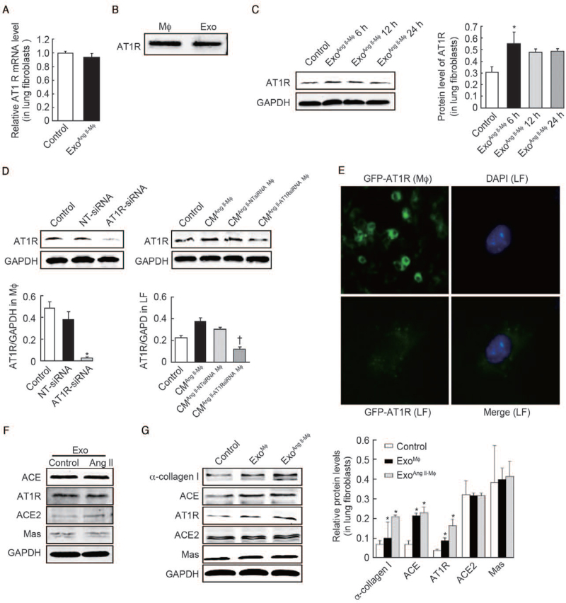
Figure 5.
AT1R-containing exosomes directly deliver AT1R to lung fibroblasts. (A) The AT1R mRNA levels of lung fibroblasts were measured using RT-PCR after treatment with macrophage exosomes. (B) Western blot analysis of AT1R protein level in macrophages and exosomes isolated from activated macrophages. (C) Fibroblasts were treated with exosomes isolated from macrophages. Then the protein level of AT1R was analyzed by western blot at different time points. n = 3 independent experiments, ∗P < 0.05 vs. control. (D) Macrophages were transfected with AT1R siRNA for 48 h, followed by 24 h exposure to 10−7 mol/L Ang II, then the cultured medium was collected for another 24 h and was used to co-culture with fibroblasts. The protein levels of AT1R in macrophages (left) and fibroblasts (right) were measured using western blot. n = 3 independent experiments, ∗P < 0.001, †P < 0.01 vs. control. (E) Fluorescence microscope analysis of macrophages expressing AT1R-green fluorescent protein (GFP) and green fluorescence was observed in lung fibroblasts (LF) when incubated with exosomes collected from overlying media of macrophages expressing AT1R-GFP. (F) Western blot analysis of ACE, AT1R, ACE2, Mas protein levels in exosomes from macrophages treated with or without Ang II. (G) Fibroblasts were treated with exosomes from macrophages treated with Ang II or left untreated. Then the protein levels of α-collagen I, ACE, AT1R, ACE2, Mas were analyzed by western blot. n = 3 independent experiments, ∗P < 0.05 vs. control. ACE: Angiotensin-converting enzyme; ACE2: Angiotensin-converting enzyme 2; Ang II: Angiotensin II; AT1R: Angiotensin II type 1 receptor; CM: Condition media; DAPI: 4-6 diamidino–2–phenylindole; Exo: Exosomes; GAPDH: Glyceraldehyde-3-3phosphate dehydrogenase; GFP: Green fluorescent protein; LF: Lung fibroblasts; Mϕ: Macrophages; mRNA: Messenger RNA; siRNA: Small interfering RNA; RT-PCR: Reverse transcription-polymerase chain reaction.

