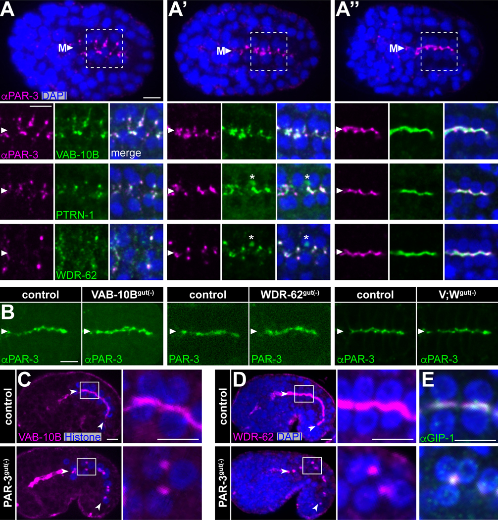
Figure 6. VAB-10B and WDR-62 interface with apical polarity determinants.
(A-A”) Dorsal views from immunofluorescence imaging of fixed embryos marking PAR-3 (anti-PAR-3, magenta), WDR-62::ZF::GFP, VAB-10B::ZF::GFP, or PTRN-1::GFP (anti-GFP, green), or DNA (DAPI, blue) from (A) pre-polarized, (A’) polarizing, and (A”) apically polarized intestines. Higher-magnification views of white boxed region in (A) shown for PAR-3 and VAB-10B and from comparable regions and stages for WDR-62 and PTRN-1. Arrowheads mark the intestinal midline (‘M’). Asterisks mark intruding green signal from ventral germ cells. (B) Immunofluorescence of fixed embryos (anti-PAR-3) or live imaging (PAR-3:mCherry) of endogenous PAR-3 in indicated genotypes. (C-E) Imaging of comma stage embryos. Higher-magnification views of white boxed region in intestinal cells are shown at right. (C) Live imaging of VAB-10B::GFP (magenta) and histone::mCherry (blue). (D, E) Immunofluorescence imaging of fixed embryos marking WDR-62::RFP (magenta) and DAPI (blue). (E) Stage-matched embryos show GIP-1 localization (anti-GIP-1, green). Scale bars = 5 μm.
See also Figures S2 and S5.

