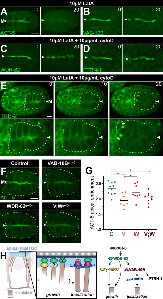
Figure 7. Actin filaments regulate non-centrosomal microtubules.
(A-D) Time-lapse live imaging of embryos expressing YFP::ACT-5 transgene (n = 3), or endogenously tagged VAB-10B::ZF::GFP (n = 4), WDR-62:ZF::GFP (n = 3) or PTRN-1::GFP (n = 3) beginning several seconds after eggshell permeabilization (t = 0’) in the presence of indicated actin inhibitors Latrunculin A (LatA) and Cytochalasin D (CytoD) for 20 minutes (t = 20’). (E) Time-lapse live imaging of GFP::TBB-2/β-tubulin with intestine (white dotted line), midline (‘M’, white triangle), and active mitotic centrosomes (asterisks) indicated; n = 3; see Video S2. (F) Live imaging of YFP::ACT-5 in control, VAB-10Bgut(−), WDR-62gut(−), or [VAB-10B; WDR-62]gut(−) (V;Wgut(−)) embryo. (G) Quantifications of apical YFP::ACT-5 fluorescent signal enrichment from (F). Each dot represents a single embryonic intestine and horizontal black lines indicate mean value; control (‘C’) n = 10; VAB-10Bgut(−) (‘V’) n = 10; WDR-62gut(−) (‘W’) n = 8; V;Wgut(−) (‘V;W’) n = 9; *p < 0.05; **p < 0.01. (H) Model of apical ncMTOC composition and function. The apical PAR complex lies upstream of divergent microtubule growth and localization modules. Scale bars = 5 μm.

