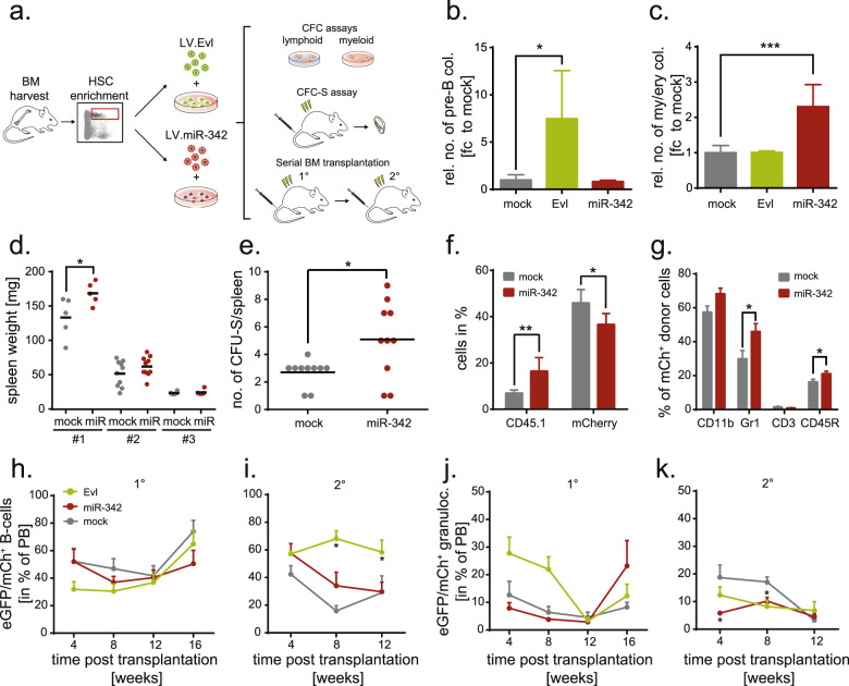
Fig. 2. EVL drives lymphopoiesis whereas miR-342 promotes myeloid differentiation in vitro and in vivo.
a Experimental setup scheme. Bone marrow (BM) was harvested from Bl6 mice, enriched for Lin− Sca1+ cKit+ (LSK) cell markers by flow cytometry and cells were transduced with overexpression (OE) lentiviral vectors for Evl or miR-342 (and mock as a control). The influence of Evl and miR-342 OE on the hematopoietic differentiation capacity was assessed by lymphoid and myeloid colony-forming assays (CFC) in vitro or a CFC-Spleen (S) assay in vivo. Multilineage reconstitution and self-renewal were addressed by serial BM transplantation. b Relative number of colonies derived from EVL-, miR-342- or mock overexpressing LSK cells in pre-B-cell growth supporting semisolid medium (n = 3). c Relative number of colonies derived from EVL-, miR-342- or mock overexpressing LSK cells in erythroid and myeloid growth supporting semisolid medium (n = 3). d Weight of spleens in mg of mice transplanted with 14,400 LSK cells/mouse (group #1), 1440 LSK cells/mouse (group #2), and 530 LSK cells/mouse (group #3) transduced with miR-342 or mock overexpression vectors 13 days post-transplant. e Number of myelopoietic progenitor-derived colony-forming units (CFU) within the spleens of mice transplanted with LSK cells OE miR-342 or mCherry control 13 days post-transplant. f Frequency of donor-derived (CD45.1) and transduced (mCherry) cells within spleens of mice at day 13 after transplantation of miR-342 OE or mock LSK cells. g Frequency of myeloid (CD11b, Gr1) and lymphoid (CD3, CD45R) hematopoietic cells within the mCherry-positive donor cell fraction in spleens of mice at day 13 after transplantation of miR-342 OE or mock LSK cells. h Frequency of donor-derived eGFP/mCherry-positive peripheral B-cells in primary (1°) and i secondary (2°) recipient mice. j Frequency of donor-derived eGFP/mCherry-positive peripheral granulocytes in primary (1°) and k secondary (2°) recipient mice. *P < 0.05; **P < 0.01; ***P < 0.001; h–k (n = 6 mice/group; SEM).

