Fig. 2.
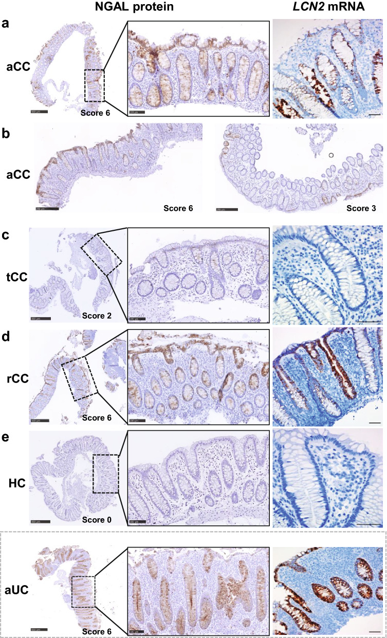
Representative images of IHC and ISH showing expression of NGAL protein and LCN2 mRNA in colonic epithelium in collagenous colitis. a Overview of NGAL protein expression (left panel), with higher magnification of epithelial staining throughout the crypts (middle panel), and LCN2 mRNA expression (right panel) in colonic mucosa of patients with active collagenous colitis (aCC). b Overview of NGAL expression in aCC mostly in the surface epithelium (left panel) or with a patchy appearance (right panel) c–e Overview of NGAL protein expression (left panel), with higher magnification as indicated (middle panel), and LCN2 mRNA expression (right panels) in colonic mucosa of patients with budesonide-treated collagenous colitis in clinical remission (tCC), d budesonide-refractory collagenous colitis (rCC) and e healthy controls (HC). Active ulcerative colitis (aUC) was included for comparison (separated by dotted grey frame). The IHC NGAL scores are given for each image. Scale bars 500 µm (left panel), 100 µm (middle panel), 50 µm (right panel)

