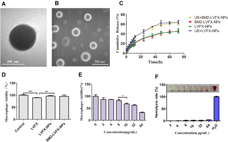
Figure 3.
Morphology and characterization of nanoparticles. (A) TEM image of BM2-LVFX-NPs. (B) SEM image of BM2-LVFX-NPs. (C) Release profiles of LVFX from drug-loaded nanoparticles with or without ultrasound exposure. (D) Cell viability of macrophages after incubation with free drugs and drug-loaded nanoparticles. (E) Cell viability of macrophages after incubation with BM2-LVFX-NPs at various LVFX concentrations. (F) Quantitative results of hemolytic activity of BM2-LVFX-NPs with different LVFX concentrations.

