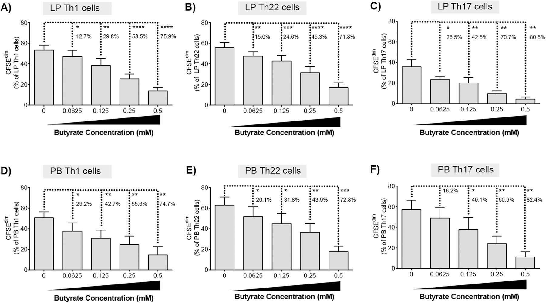
Fig. 2.

Butyrate decreases proliferation of LP and PB T helper cells. CFSE-labelled (A) LPMC (N = 6) or (B) PBMC (N = 6) were cultured with TCR-activating beads and exogenous butyrate (0.0625–0.5 mM) for four days with mitogenic stimulation during the last 4 h of culture. Percentages of proliferating (A, D) Th1, (B, E) Th22 or (C, F) Th17 cells were determined using multi-color flow cytometry. Isotype control values have been subtracted. Values are shown as mean ± SEM of net proliferation (TCR-stimulated minus unstimulated). Statistical analysis: Paired t tests were conducted to determine differences in Th subset proliferation between butyrate concentrations versus no butyrate conditions. Percentages represent the average percentage decrease in Th cell proliferation versus no butyrate. *P < 0.05, **P < 0.01, ***P < 0.001, ****P < 0.0001.
