
Figure 4. VM axon activation facilitates lick initiation by activating ALM.
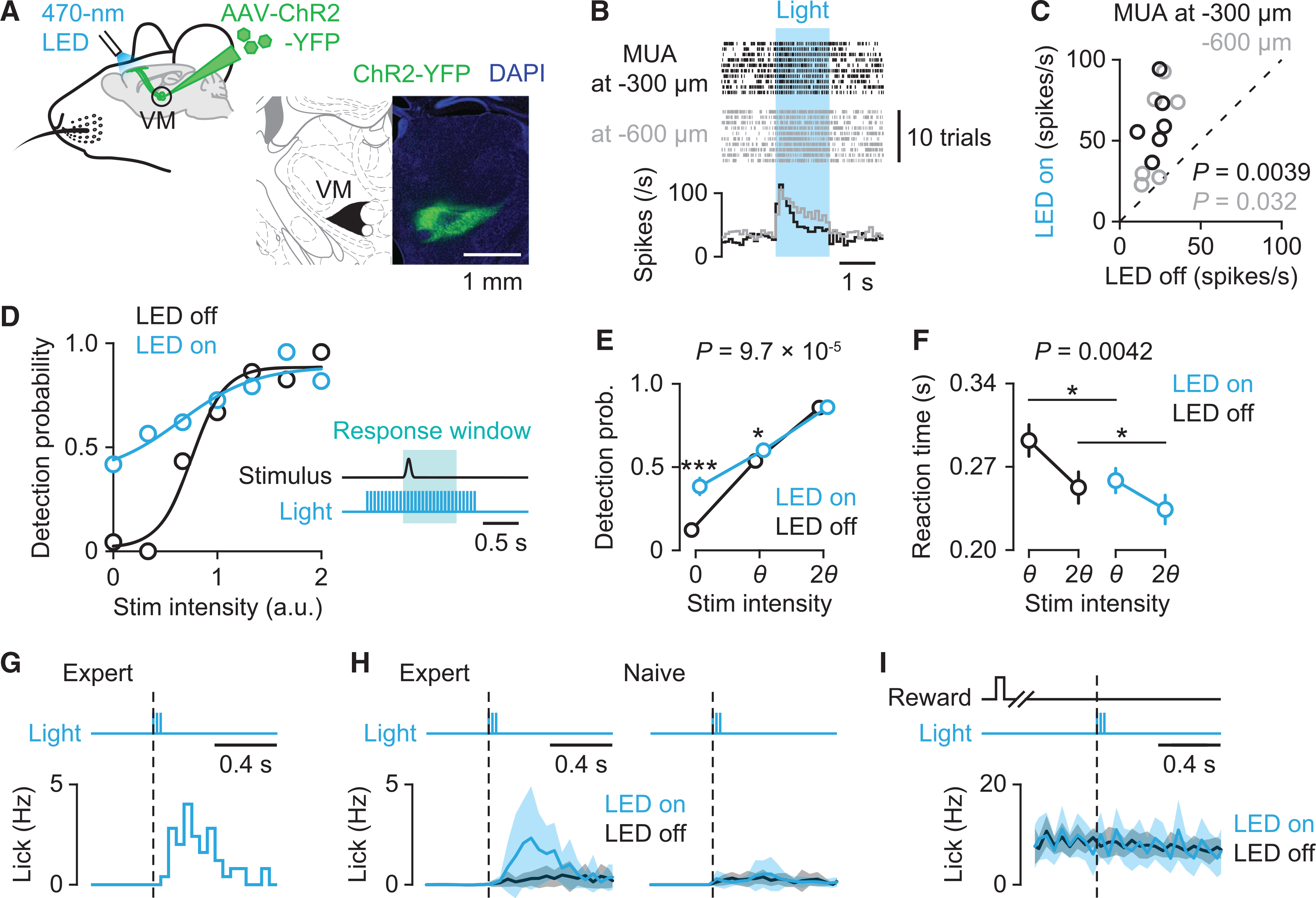
(A) Left: Schematic showing optogenetic stimulation of ChR2-expressing VM axons in ALM. Right: ChR2-YFP expression in VM.
(B) Raster plot (top) and peri-stimulus time histogram (PSTH) (bottom) of multi-unit activity (MUA) in ALM at 300 μm (black) and 600 μm (gray) below the pia during photostimulation (blue).
(C) Relationship between MUAs during control (LED off) and photostimulation (LED on) conditions (n = 6 recording sites for each depth from four mice; paired t test).
(D) Example of psychometric performance with (blue) or without (black) photostimulation of VM axons. Inset: Schematic showing the time course of photostimulation. 470 nm light (5 ms duration at 20 Hz) was delivered to the left ALM.
(E and F) Behavioral comparisons with (blue) and without (black) photostimulation. (E) Detection probability (n = 16 sessions from four mice, mean ± SEM; two-way repeated-measure ANOVA with Tukey–Kramer post hoc comparisons, ***p = 4.5 × 10⁻⁵ for 0, *p = 0.037 for θ). (F) Reaction times for threshold and salient stimuli (n = 16 sessions from four mice, mean ± SEM; two-way repeated-measure ANOVA with Tukey–Kramer post hoc comparisons, *p = 0.012 for θ; *p = 0.036 for 2θ).
(G) Lick responses to brief photostimulation (three pulses at 20 Hz) in catch trials in a representative session.
(H) Lick responses to brief photostimulation averaged across sessions in trained expert mice (n = 10 sessions from five mice, mean ± SEM; left) and untrained naive mice (n = 10 sessions from five mice, mean ± SEM; right).
(I) Impact of brief photostimulation during ongoing licking in hit trials in expert mice (n = 10 sessions from five mice, mean ± SEM).
See also Figure S4.