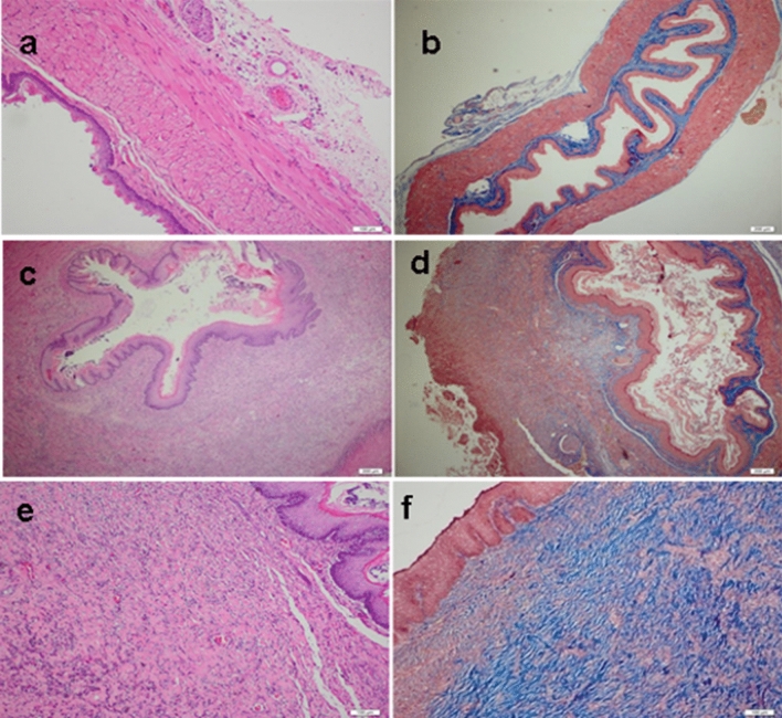
Figure 2.
Mean histopathological pictogrammes of the groups studied. (a, b) Esophageal mucosa belonging to the control group. (a) There is normal archetype preserved esophageal full-thickness mucosa sample containing lamina propria and muscularis propria consisting of compact connective tissue observed under the surface ceratinized squamous epithelium, in the Hematoxylin&Eosin (H&E), × 100). (b) There is no collagen increase in the submucosal and muscular layers, in the Mason Trichrome Stain, × 100. (c, d) Esophageal mucosa belonging to the groups of sham and FGF(−). For the Groups of Sham and FGF(−) not received FGF, there are noteworthy epithelial degeneration, intense inflammation in the submucosa and muscular layer, and mildly increased collagen, in the Mason Trichrome Stain, × 100 and Hematoxylin&Eosin (H&E), × 100. (e, f) Esophageal mucosa belonging to the groups given FGF. (e) In the surface epithelium, there is marked regeneration as well as the presence of significantly increased collagen, which is replaced by the submucosal and muscular layer. There is a moderate decrease in the inflammatory cell density observed between the collagen bundles on the 7th day and a significant decrease on the 28th day, in the Hematoxylin&Eosin (H&E), × 100. (f) There is significantly increased collagen (blue color) between the submucosal and muscular layers observed, in the Mason Trichrome Stain, × 100.

