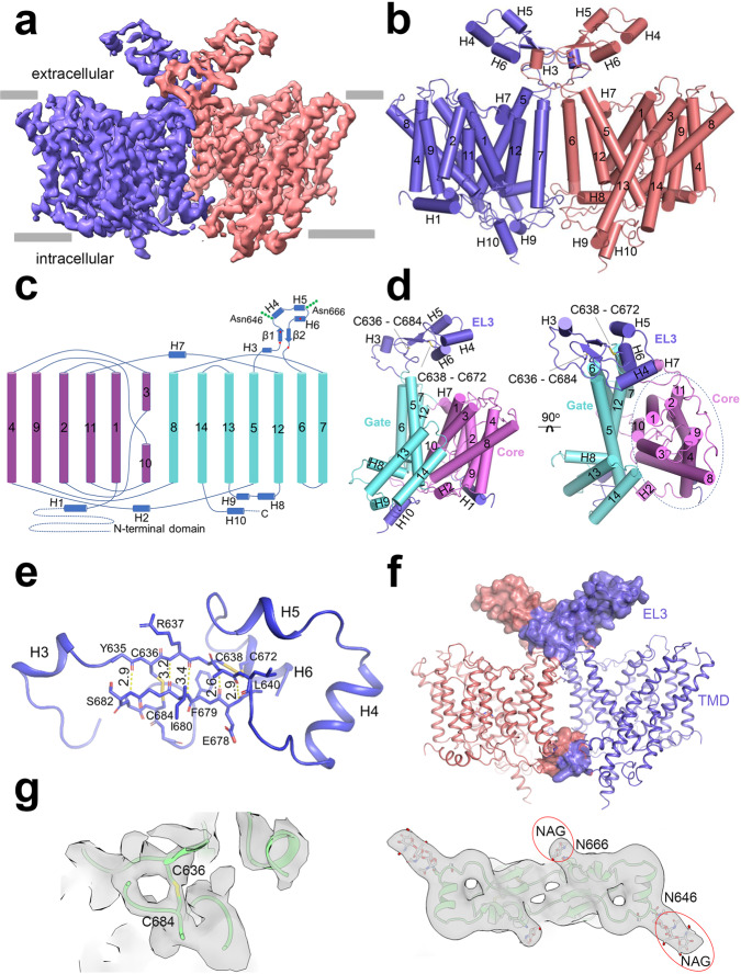
Fig. 1. Overall structure of NDCBE TMD.
a Side view of a three-dimensional (3D) reconstruction of NDCBE with individually colored monomers. b Cartoon representation of NDCBE dimer. TMs 1-14 and helices H1-10 are shown as cylinders. c Topology and domain arrangement of the NDCBE monomer. Four highly conserved cysteines are labeled as red dot. The branched structures at Asn646 and Asn666 represent N-linked glycosylation. d Structure of NDCBE monomer in side (left) and extracellular (right) views. e Interactions contributing to EL3 assembly. f EL3 and TMD components involved in formation of the dimeric interface. g Cryo-EM densities showing the disulfide bond between Cys636 and Cys684 (left) and two N-glycosylation sites at Asn646 and Asn666 (right); the map was low-pass filtered to 5 Å for the two N-glycosylation sites.

