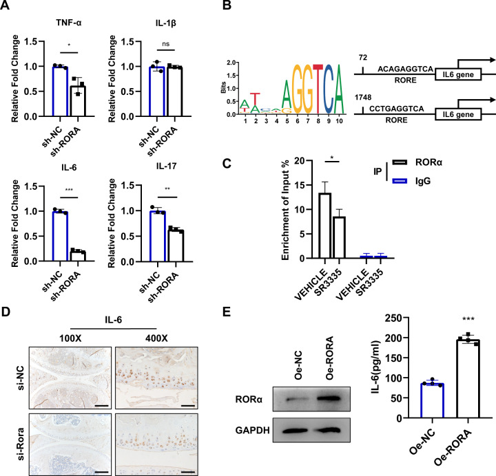
Fig. 7. RORα regulates IL-6 expression by interacting with IL-6 promoters.
A Real-time PCR analysis of TNF-α, IL-1β, IL-6, and IL-17 mRNA in human chondrocytes infected with RORA knockdown lentivirus (sh-RORA) or control lentivirus (sh-NC) (n = 3). B Schematic diagram of the potential binding site for RORα in the promoter region of IL-6 using JASPAR database. C ChIP-qPCR enrichment assay was performed on chondrocytes with anti-RORα antibody or IgG. The chondrocytes were pretreated with vehicle or 1 μM SR3335 for 6 h. IgG immunoprecipitation was used as a negative control. D IHC staining for IL-6 of articular cartilage after indicated adenovirus injection for 6 weeks in mice. Scale bars, 200 μm for ×100 picture and 50 μm for ×400 picture. E ELISA assay to detect IL-6 protein expression after transfected with RORA overexpression or vector plasmid for 48 h (n = 4). The statistical data in A, C, and E were analyzed with Student’s t test. *P < 0.05, **P < 0.01, ***P < 0.001, ns = no significance. All data shown above are presented as mean ± SD.

