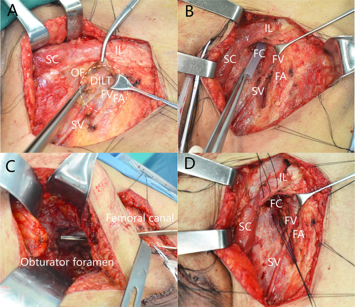
Figure 1.
Deep inguinal lymph nodes dissection. (A) Position of deep inguinal lymph nodes. (B) The femoral canal is empty after removal of DILT. (C) Femoral canal communicates with obturator after removal of DILT and pelvic LNs. (D) Closing the femoral canal. DILT, deep inguinal lymphatic tissue; FA, femoral artery; FC, femoral canal; FV, femoral vein; IL, inguinal ligament; OF, oval fossa; SC, spermatic cord; SV, saphenous vein.

