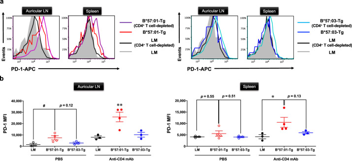
Fig. 2. PD-1 surface expression of effector memory CD8+ T cells in CD4+ T cell-depleted HLA-transgenic (Tg) mice.
a Flow cytometric measurement of PD-1 surface expression. Representative histograms of PD-1 surface expression on effector memory CD8+ T cells in the auricular lymph node (LN) or spleen from CD4+ T cell-depleted/PBS-treated B*57:01-Tg mice (left panel), B*57:03-Tg mice (right panel), or their littermates (LMs). Mice have orally administrated 1% (w/w) abacavir (ABC) for 1 week. Effector memory CD8+ T cells were gated from either lymphocytes or splenocytes by anti-CD44 and CD62L antibodies (phenotype: CD44highCD62Llow). Data are representative of three independent experiments. b Median fluorescence intensity (MFI) values of PD-1 in effector memory CD8+ T cells isolated from the auricular LN (left panel) or spleen (right panel) of 1% (w/w) ABC-fed mice for 1 week. Each plot represents an individual mouse with the mean ± SEM (N = 3–4); #p < 0.05, one-way ANOVA with Bonferroni’s multiple comparisons correction, compared with PBS-treated mice groups; indicated p values were obtained from a statistical comparison. *p < 0.05, **p < 0.01, one-way ANOVA with Bonferroni’s multiple comparisons correction, compared with other mice groups. Data are a summary of three independent experiments.

