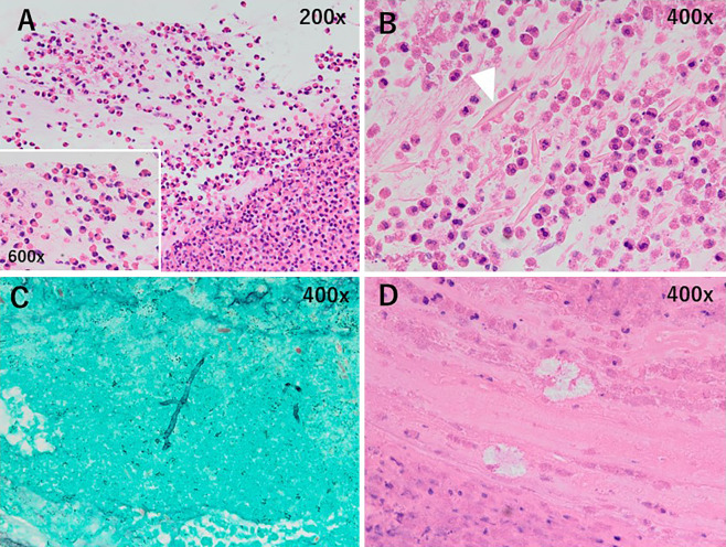
Figure 4.
Hematoxylin and Eosin staining shows that the material biopsied from the mucoid impaction contains abundant eosinophils (A, 200×, inset 600×), Charcot-Leyden crystals (B, 400×, arrowhead), and calcium oxalate crystals (D, 400×). Filamentous fungal hyphae are noted on the Papanicolaou-stained specimen (C, 400×).

