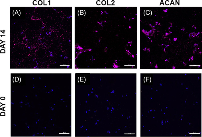
FIGURE 8.

Representative immunofluorescent staining (magenta) of A, COL1; B, COL2, and C, ACAN produced by adipose derived mesenchymal stem cells (ADMSCs) cultured within formulation S‐50 for 14 days in the presence of soluble GDF‐6. Staining for day 0, immediately after encapsulation, is presented as a comparison in D‐F. Cell nuclei are counterstained with DAPI (blue). Scale bars = 100 μm
