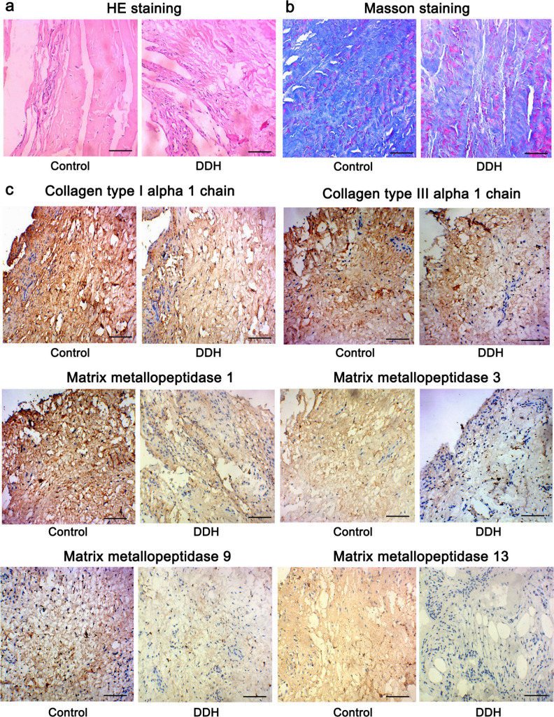
Fig. 2.
Histological examinations using haematoxylin and eosin (HE), Masson’s, and immunohistochemistry staining (200×). Tissues from the hip joint capsule from patients with developmental dysplasia of the hip (DDH) and healthy controls were subjected to a) HE, b) Masson’s, and c) immunohistochemistry staining. Bar: 10 μm.

