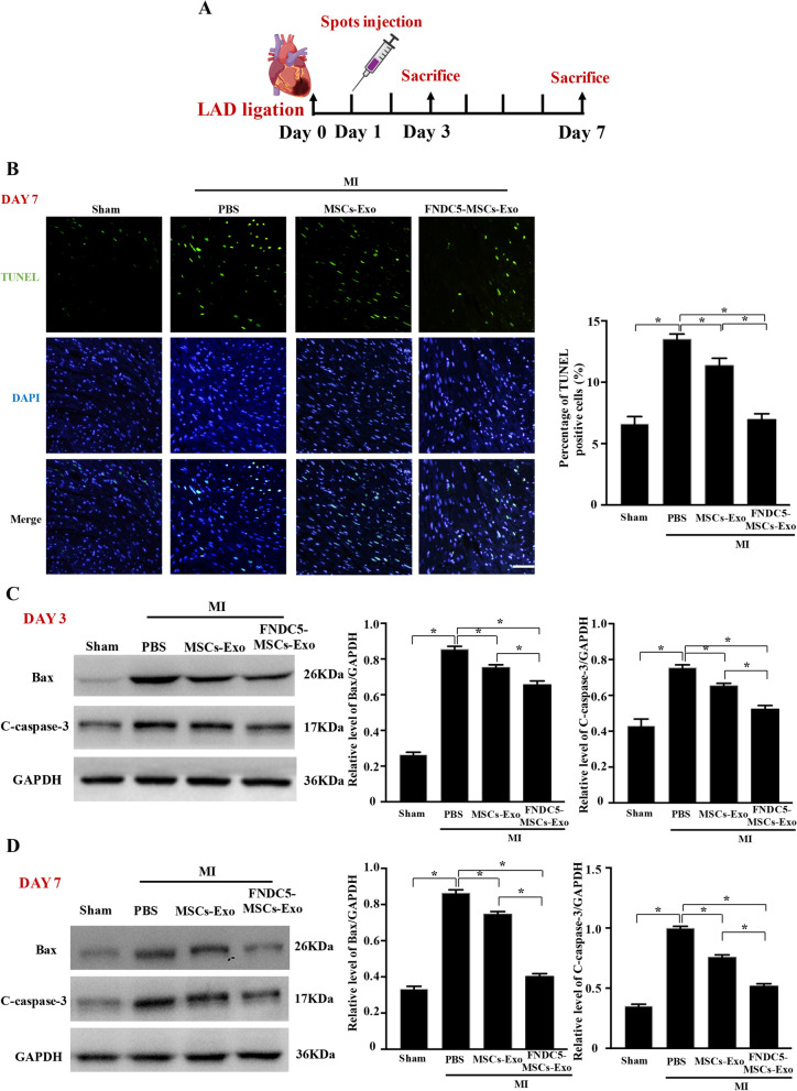
Fig. 1.
Intramyocardial spots injection of exosomes derived from FNDC5-MSCs significantly suppressed cardiomyocyte apoptosis in MI mice. a The diagrammatic sketch of MI induction and exosome intramyocardial injection and mice were killed at 3 and 7 days after intramyocardial injection (n ≥ 14). b The apoptosis of cardiomyocytes at 7 days was assessed by a TUNEL Assay Kit (scale bars = 20 μm). Nucleus was blue with DAPI and the TUNEL-positive apoptotic cardiomyocytes were green. The average fluorescence intensity of positive apoptotic cardiomyocytes was quantified. The protein of Bax and cleaved caspase‐3 within the heart tissue at 3 days (c) and 7 days (d) was detected by Western blot and semi-quantification analysis. Data are expressed as the means ± SEM; n = 5; *P < 0.05

