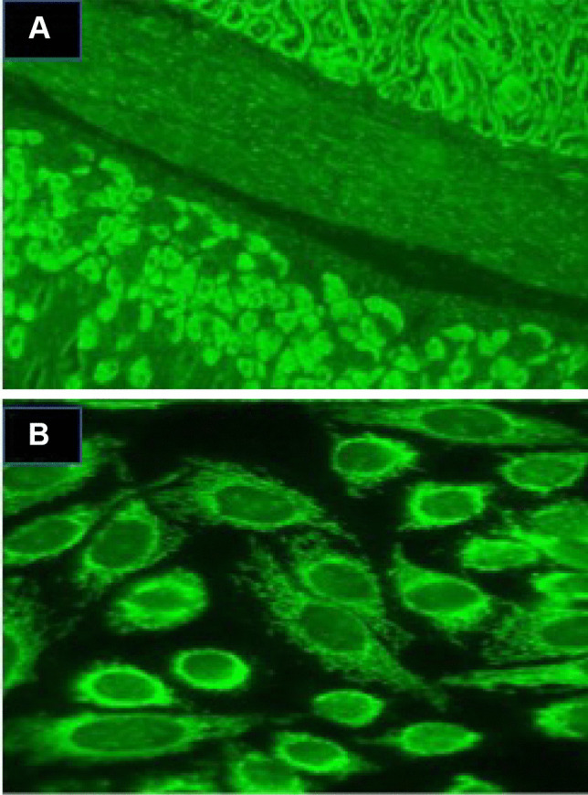
Fig. 1.

Anti-mitochondrial antibodies detection by indirect immunofluorescence using HEp-2 cell or mouse tissue sections. A AMA staining of mouse kidney/smooth muscle/stomach tissue section showing staining of both proximal and distal tubules of the mouse kidney (upper right) and the parietal cells of the mouse stomach (lower left). B AMA on HEp-2 cells
