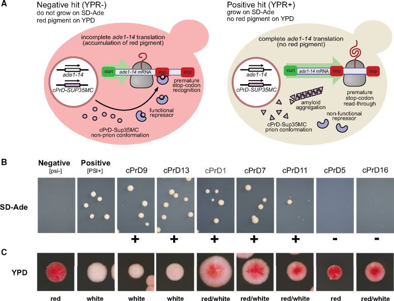
Fig. 6.
cPrD-Sup35MC strains display different ability to grow on media lacking adenine and a variety of colony colors. (A) Cartoon explaining the mechanism of accumulation of red pigment, which depends on the formation of functional repressor. (B and C) Representative images of the colonies of cPrD-Sup35MC-expressing strains growing on SD-Ade and YPD plates respectively (see supplementary fig. S5, Supplementary Material online, for all 16 tested and supplementary fig. S7, Supplementary Material online, for images of the whole area of Petri dishes). Colonies phenotypes [psi−] and [PSI+] are shown for comparison. Positive results are marked with “+” and are considered positive results of the yeast prion reporter assay (YPR+). Negative results are marked with “−” and are considered negative results of the yeast prion.

