Figure 5.
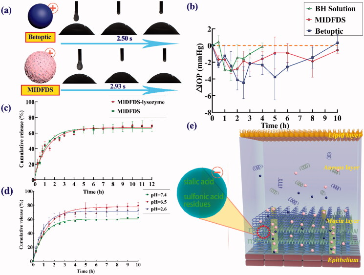
Pharmacodynamics and corneal preparation spread time. (a) The spreading time of Betoptic and MIDFDS at the front of the rabbit cornea in vitro. (b) IOP-lowering effects of topical administration of BH solution, Betoptic, and MIDFDS (mean ± SD, n = 3). (c) In vitro release of BH from MIDFDS and MIDFDS-lysozyme (n = 3). (d) In vitro release of BH from MIDFDS at different pH values (n = 3). (e) Schematic diagram of the micro-interaction between the positively charged microspheres and the negatively charged mucin layer after local administration. The larger corneal contact angle of MIDFDS and the slower corneal diffusion velocity in rabbit eyes led to increased interaction time between the positively charged surface of MIDFDS and the negative residues of the tear film, further resulting in a longer precorneal retention time of MIDFDS in in vivo fluorescence tracing (Figure 4(a)).

