. 2021 Sep 2;12(37):12266–12273. doi: 10.1039/d1sc01784e

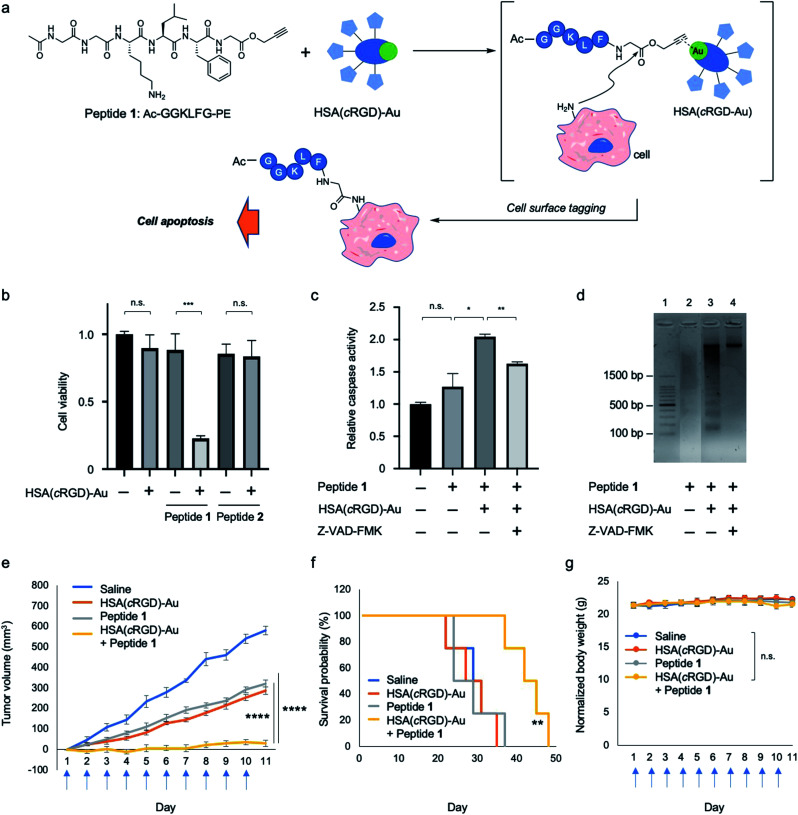
Fig. 3. Cell surface modification with proapoptotic peptide 1 with propargyl ester attached at the C-terminus by Au-catalyzed amide bond formation (Au/PE). (a) Cell surface modification with proapoptotic peptide 1 and HSA(cRGD)-Au. (b) Cytotoxicity of peptide 1 or peptide 2 (600 μM) with or without HSA(cRGD)-Au (20 μM) in SW620 cells (n = 3, mean ± SE). (c) Caspase activities (n = 3, mean ± SE) in the presence of caspase inhibitor (Z-VAD-FMK). (d) Electrophoresis image of DNA fragmentation in cells treated with peptide 1 cells with or without HSA(cRGD)-Au. Effects of tumor therapy on (e) tumor volume, (f) survival probability, and (g) body weight changes of SW620-xenografted mice subjected to the following treatments: saline (n = 4, blue), HSA(cRGD)-Au (35.0 mg kg−1, n = 7, orange), peptide 1 (4.0 mg kg−1, n = 7, gray), and co-treatment of peptide 1 and HSA (cRGD)-Au (4.0 mg kg−1 and 35.0 mg kg−1, n = 7, yellow). Blue arrows under the horizontal axis indicate the day of treatment with compounds. Data are shown as means ± SE. n.s.: not significant, *p < 0.05, **p < 0.01, ***p < 0.005, ****p < 0.001.

Fig. 3